Amorphous


Inquiry number
SOL-0000001137
Beamline
BL04B2 (High Energy X-ray Diffraction)
Scientific keywords
| A. Sample category | inorganic material |
|---|---|
| B. Sample category (detail) | amorphous, glass |
| C. Technique | X-ray diffraction |
| D. Technique (detail) | wide angle scattering |
| E. Particular condition | room temperature |
| F. Photon energy | X-ray (> 40 keV) |
| G. Target information | structure analysis |
Industrial keywords
| level 1---Application area | cell (battery), industrial material, others |
|---|---|
| level 2---Target | fuel cell |
| level 3---Target (detail) | |
| level 4---Obtainable information | structure of non-crystalline material |
| level 5---Technique | diffraction |
Classification
M20.20 middle angle scattering
Body text
High-energy x-ray diffraction is a powerful technique to study the structure of amorphous materials. Using this technique, one can obtain atomic correlations of disordered structure. The high-flux high-energy x-rays allow us to measure reliable diffraction pattern of amorphous materials.
Figures 1 (a) and (b) show the structure factors S(Q) of HoFe2D3.0 and TbFe2D3.0, respectively. Figures 1 (c) and (d) show corresponding atomic configurations derived from computer simulation based on diffraction data. As can be seen from these atomic configurations, The concentration fluctuations between the metal atoms are clearly visible in both systems. In addition, many deuterium atoms are located in the clusters of rare-earth metals.
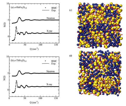
Fig. 1 The structure factors S(Q) of HoFe2D3.0 (a) and TbFe2D3.0 (b), and atomic configurations of HoFe2D3.0 (c) and TbFe2D3.0 (d).
[ K. Itoh, Y. Miyajima, K. Aoki and T. Fukunaga, Journal of Alloys and Compounds 376, 9-16 (2004), Fig. 5, 8,
©2004 Elsevier Science Publisher ]
Source of the figure
Original paper/Journal article
Journal title
J. Alloys Comp., 376, 9-16 (2004).
Figure No.
5,8
Technique
High-energy x-ray diffraction is a powerful tool to study the structure of disordered materials. The analysis of the diffraction pattern makes it possible to derive inter-atomic distance, coordination number and so on. One of the advantage of the diffraction in comparison with EXAFS is that intermediate-range order, which can be analyzed by computer simulation based on diffraction data. A combination of high-energy x-ray diffraction, neutron diffraction, and computer simulation is an essential tool to study the structure of disordered materials.
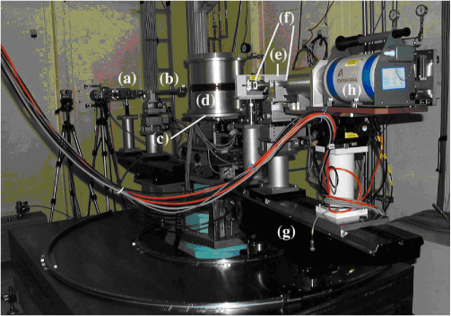
[ S. Kohara, Y. Ohishi, M. Takata, Y. Yoneda and K. Suzuya, Journal of the Crystallographic Society of Japan 47, 123-129 (2005), Fig. 1,
©2005 The Crystallographic Society of Japan ]
Source of the figure
Original paper/Journal article
Journal title
日本結晶学会誌,47,123-129(2005)
Figure No.
Required time for experimental setup
4 hour(s)
Instruments
| Instrument | Purpose | Performance |
|---|---|---|
| Two-axis diffractometer | to get diffraction pattern | 113.4 keV |
References
| Document name |
|---|
| X線構造解析—原子の配列を決める 材料学シリーズ 早稲田 嘉夫, 松原 英一郎 |
Related experimental techniques
Neutron diffraction
Questionnaire
The measurement was possible only in SPring-8. Impossible or very difficult in other facilities.
This solution is an application of a main instrument of the beamline.
Similar experiments account for more than 30% of the beamline's subject.
Ease of measurement
Easy
Ease of analysis
Middle
How many shifts were needed for taking whole data in the figure?
Two-three shifts