In-situ observation of growth of Fe-Zn intermetallic compounds in galvannealing process

Inquiry number
SOL-0000000881
Beamline
BL19B2 (X-ray Diffraction and Scattering II)
Scientific keywords
| A. Sample category | inorganic material |
|---|---|
| B. Sample category (detail) | metal, alloy |
| C. Technique | X-ray diffraction |
| D. Technique (detail) | wide angle scattering |
| E. Particular condition | high-T (~500 C), time-resolved (slow) |
| F. Photon energy | X-ray (4-40 keV) |
| G. Target information | structural change |
Industrial keywords
| level 1---Application area | mechanics, construction, industrial material |
|---|---|
| level 2---Target | Steel |
| level 3---Target (detail) | film, lubricant |
| level 4---Obtainable information | surface,interface, crystal structure, orientation (preferred orientation) |
| level 5---Technique | diffraction |
Classification
A80.20 metal ・material
Body text
In this solution, in-situ X-ray diffraction measurement was applied to analyze the growth behavior of Fe-Zn intermetallic compounds at initial stage (~ a few 10 seconds) of gaalvannealing process. The data of change of the X-ray diffraction pattern from zinc coating indicated the dependence of the composition of the coatings on the growth behavior of Fe-Zn intermetallic compounds in the galvannealing process.
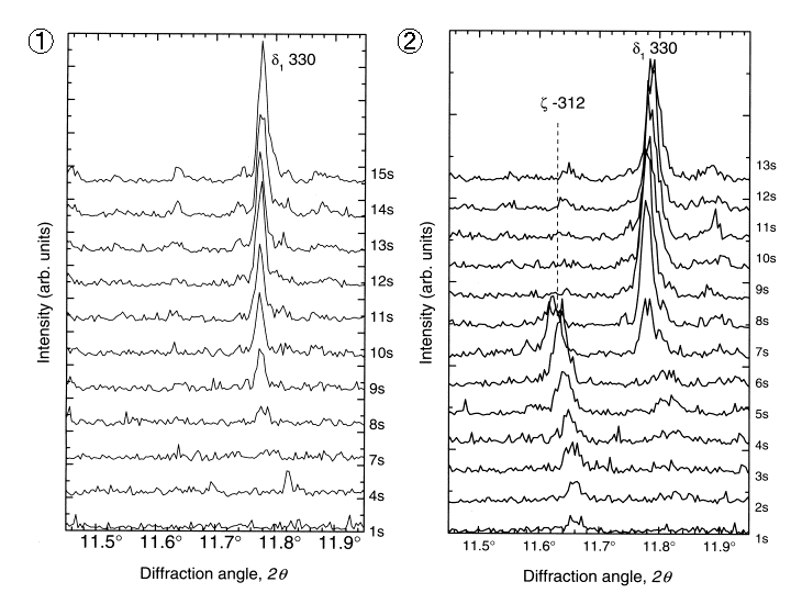
Change of the X-ray diffraction profiles from the Zinc coating in the galvannealing process
1.Zn coating with 0.13mass% Al 2.Pure Zn coating
[ A. Taniyama, M. Arai, T. Takayama and M. Sato, Materials Transactions 45, 2326-2331 (2004), Fig. 4, 7,
©2004 The Japan Institute of Metals ]
Source of the figure
Original paper/Journal article
Journal title
A. Taniyama, M.Arai, T. Takayama and M. Sato, Material ransaction Vol.45, No.7 (2004) pp.2326-2331
Figure No.
Fig.4,Fig.7
Technique
In this experiment, the measurement of the X-ray diffraction from galvanized steel were carried out heating the samples up to T = ~460C by infrared beam heater. The time dependence of 1D profiles of the diffraction extracted by a receiving slit (2.5400mm) were continuously recorded on imaging plates set behind the receiving slit scanning in the horizontal direction. Using high energy X-ray (28KeV), the initial stage (~10sec)of the growth behavior of Fe-Zn intermetallic compounds at the interfaces between the Zn coatings and substrates was successfully observed.
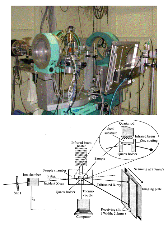
The photograph and the schematic view of the experimental instrument
[ A. Taniyama, M. Arai, T. Takayama and M. Sato, Materials Transactions 45, 2326-2331 (2004), Fig. 1,
©2004 The Japan Institute of Metals ]
Source of the figure
Original paper/Journal article
Journal title
A. Taniyama, M. Arai, T. Takayama and M. Sato, Material Transactions, Vol. 45, No. 7 (2004) pp.2326-2331
Figure No.
Fig.1
Required time for experimental setup
9 shift(s)
Instruments
| Instrument | Purpose | Performance |
|---|---|---|
| Multi-axis diffractometer | Measurement of the X-ray diffraction | Camera length for IP camera : 40~80cm |
| IP camera | Recording of the X-ray diffraction profile | Area of IP : 200×400mm |
References
| Document name |
|---|
| Akira Taniyama, Masahiro Arai, Toru Takayama and Masugu Sato, Material Tranzaction, Vol. 45, no. 7 (2004) p2326-2331 |
Related experimental techniques
Questionnaire
The measurement was possible only in SPring-8. Impossible or very difficult in other facilities.
This solution is an application of a main instrument of the beamline.
With user's own instruments.
Ease of measurement
With a great skill
Ease of analysis
Middle
How many shifts were needed for taking whole data in the figure?
Four-nine shifts