DEI movie


問い合わせ番号
SOL-0000001150
ビームライン
BL20B2(医学・イメージングI)
学術利用キーワード
| A. 試料 | 生物・医学, 計測法、装置に関する研究 |
|---|---|
| B. 試料詳細 | 低分子有機材料, 高分子有機材料, 生体(in vivo), 生体(in vitro), 生体組織、細胞系等, 生体材料, 生体高分子、結晶, 生体高分子、非結晶 |
| C. 手法 | X線回折, 吸収、及びその二次過程 |
| D. 手法の詳細 | 反射、屈折 |
| E. 付加的測定条件 | 二次元画像計測, 界面, 室温 |
| F. エネルギー領域 | X線(4~40 keV), X線(>40 keV) |
| G. 目的・欲しい情報 | 形態・巨視的構造 |
産業利用キーワード
| 階層1 | 製薬, その他 |
|---|---|
| 階層2 | 製剤 |
| 階層3 | 薬物 |
| 階層4 | 密度, 亀裂、空隙, 形態 |
| 階層5 | イメージング |
分類
A80.50 製薬
利用事例本文
DEI (Diffraction Enhanced Imaging) is a unique technique to obtain an image of an object based on the difference in the refractive indices. The technique gives an image with a unique contrast which provides information on the fine structure of the object. By using a CMOS flatpanel detector, it is possible to observe an object continuously at a frame rate of up to 2 per second. The figure reveals movement of mouse knee joint with unprecedented contrast.
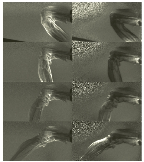
[ K. K. W. Siu, M. J. Kitchen, K. M. Pavlov, J. E. Gillam, R. A. Lewis, K. Uesugi and N. Yagi, Nuclear Instruments and Methods in Physics Research A 548, 169-174 (2005), Fig. 4,
©2005 Elsevier Science Publisher ]
画像ファイルの出典
原著論文/解説記事
誌名
Nuclear Instruments and Methods in Physics Research A 548 (2005) 169–174
図番号
4
測定手法
画像ファイルの出典
図なし
測定準備に必要なおおよその時間
1 シフト
測定装置
参考文献
| 文献名 |
|---|
| Nuclear Instruments and Methods in Physics Research A 548 (2005) 169–174 |
関連する手法
アンケート
SPring-8だからできた測定。他の施設では不可能もしくは難しい
本ビームラインの主力装置を使っている
測定の難易度
初心者でもOK
データ解析の難易度
初心者でもOK
図に示した全てのデータを取るのにかかったシフト数
1シフト以下