血管造影による腫瘍増殖過程の観察

Inquiry number
SOL-0000001443
Beamline
BL20B2 (Medical and Imaging I)
Scientific keywords
| A. Sample category | biology, medicine |
|---|---|
| B. Sample category (detail) | biology (in vivo) |
| C. Technique | absorption and its secondary process |
| D. Technique (detail) | |
| E. Particular condition | 2D imaging, time-resolved (ms) |
| F. Photon energy | X-ray (4-40 keV) |
| G. Target information | morphology |
Industrial keywords
| level 1---Application area | Pharmaceuticals, others |
|---|---|
| level 2---Target | process analytical technology (PAT) |
| level 3---Target (detail) | drug, organism |
| level 4---Obtainable information | molphology |
| level 5---Technique | imaging |
Classification
A80.90 others
Body text
体重3~4kgのウサギの耳介へ腫瘍細胞を移植し、1~7日経過した後での腫瘍の増殖過程を、微小血管造影で観察し、この結果を図1にまとめました。図の下段は上段の画像と同じであり点線が、腫瘍の大きさと位置を示しています。従来にない微小な血管構築の画像化が可能となり、前臨床試験へ適用すれば、早期診断や新しい癌治療法の研究のための有効な手段となるであろうと考えられます。
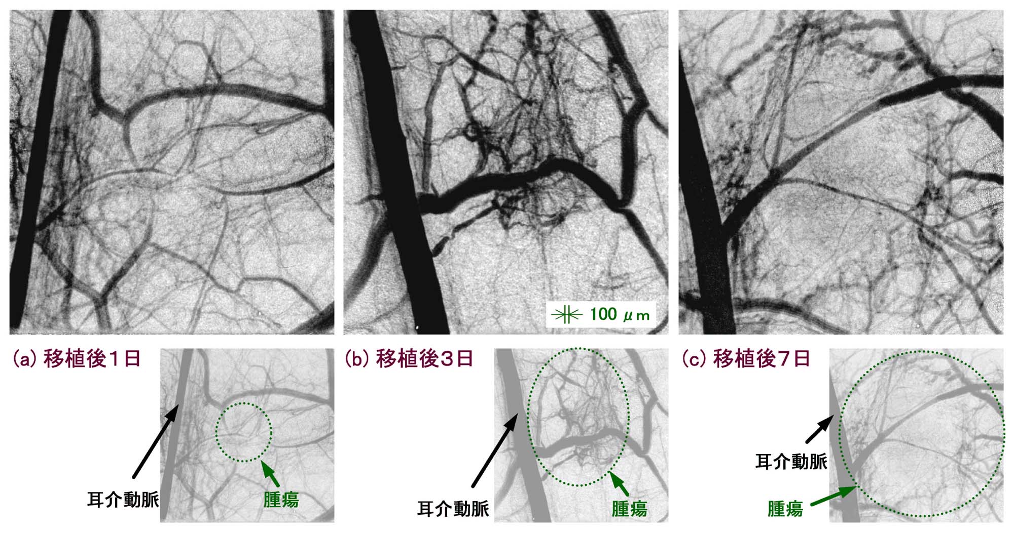
図1 ウサギ耳介腫瘍の移植後1~7日での撮影結果
[ K. Umetani, T. Yamashita, N. Maehara and S. Imai, 映像情報メディア学会誌 56, 492-494 (2002), Fig. 3,
©2002 映像情報メディア学会 ]
Source of the figure
Original paper/Journal article
Journal title
Journal of Institute of Image Information and Television Engineers 56(3) 492-494 (2002)
Figure No.
3
Technique
微小血管造影のため、ハイビジョン級で走査線数1050本の、X線直接変換型撮像管カメラを用いた解像度6~10µmの撮影装置を開発しました。そして、腫瘍移植ウサギの実験で、解像度10µmのモードでの撮影で20~30µm径の腫瘍血管が画像化でき、悪性腫瘍に伴う微小血管の動態観察が可能となりました。
Source of the figure
No figure
Required time for experimental setup
3 hour(s)
Instruments
| Instrument | Purpose | Performance |
|---|---|---|
| 血管造影装置 | 微小血管造影 | 解像度6~10μm |
References
| Document name |
|---|
| 映像情報メディア学会誌 56(3) 492-494 (2002) |
Related experimental techniques
特になし
Questionnaire
The measurement was possible only in SPring-8. Impossible or very difficult in other facilities.
Ease of measurement
Middle
Ease of analysis
Middle
How many shifts were needed for taking whole data in the figure?
Two-three shifts