Angiographic observation of tumor growth

Inquiry number
SOL-0000001145
Beamline
BL20B2 (Medical and Imaging I)
Scientific keywords
| A. Sample category | biology, medicine |
|---|---|
| B. Sample category (detail) | biology (in vivo) |
| C. Technique | absorption and its secondary process |
| D. Technique (detail) | |
| E. Particular condition | 2D imaging, time-resolved (ms) |
| F. Photon energy | X-ray (4-40 keV) |
| G. Target information | morphology |
Industrial keywords
| level 1---Application area | Pharmaceuticals, others |
|---|---|
| level 2---Target | process analytical technology (PAT) |
| level 3---Target (detail) | drug, organism |
| level 4---Obtainable information | molphology |
| level 5---Technique | imaging |
Classification
A80.90 others
Body text
Microangiography with spatial resolution in the micrometer range has been carried out for depicting the microangioarchitecture of tumor-derived angiogenic vessels in a rabbit model of cancer using a high-resolution detector. A VX2 carcinoma was transplanted into a rabbit auricle. Small tumor blood vessels in an immature vascular network produced by angiogenesis were visualized after contrast agent injection. Images at 1, 3 and 7 days after transplantation are shown in Fig. 1. The tumor blood vessels exhibit a characteristic appearance, and small blood vessels of 20-30 µm diameter are depicted in these images.
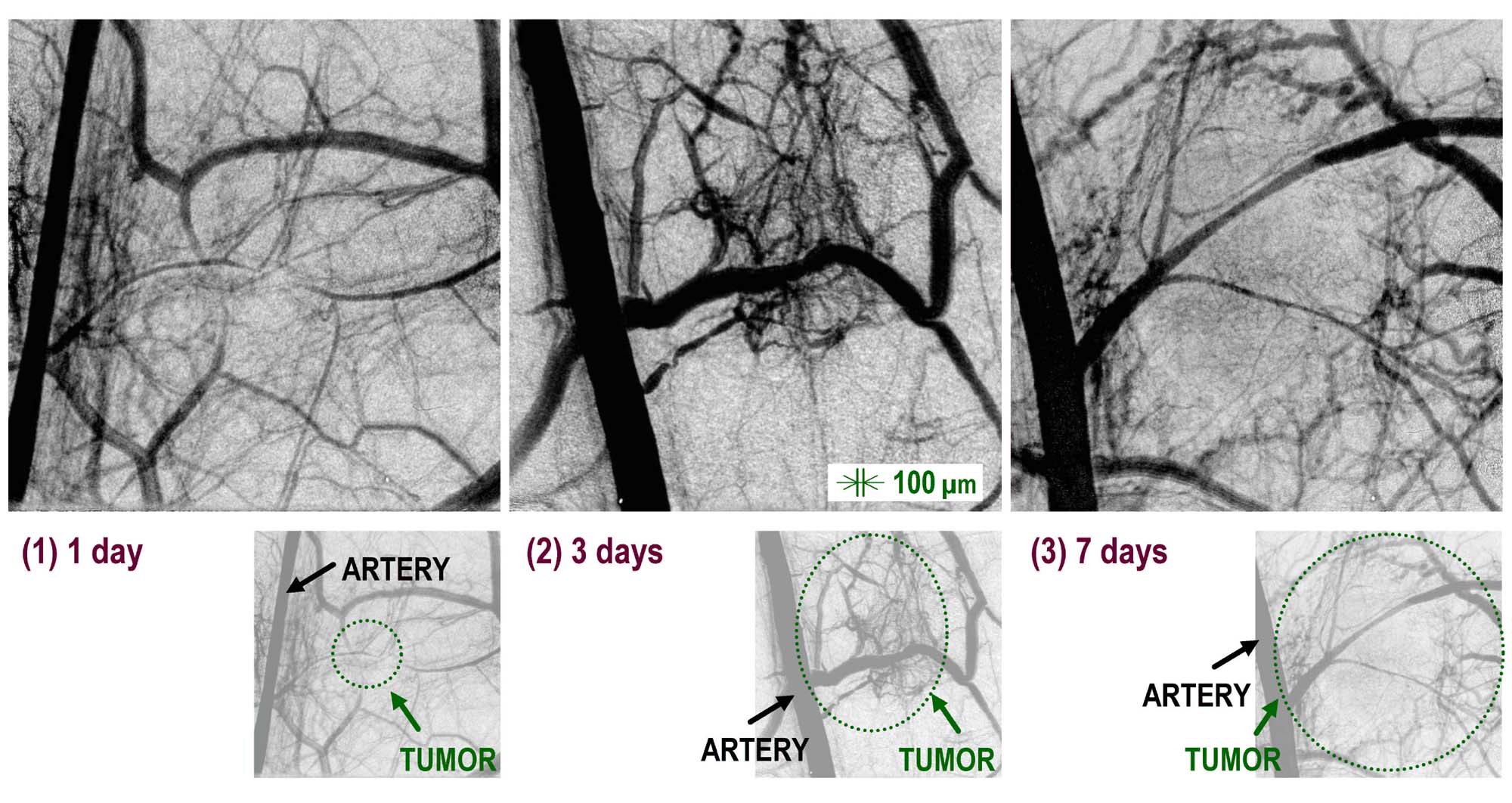
Fig. 1 Angiographic images of tumor blood vessels at 1, 3 and 7 d after tumor transplantation.
[ K. Umetani, T. Yamashita, N. Maehara and S. Imai, Journal of Institute of Image Information and Television Engineers 56, 492-494 (2002), Fig. 3,
©2002 The Institute of Image Information and Television Engineers ]
Source of the figure
Original paper/Journal article
Journal title
Journal of Institute of Image Information and Television Engineers 56(3) 492-494 (2002)
Figure No.
3
Technique
The imaging system was composed of an X-ray direct-sensing type detector incorporating an X-ray SATICON pickup tube. Angiographic images were obtained and stored in a digital frame memory system with a 1024 x 1024-pixel, 10-bit format. Sequential images are obtained with an input field of 9.5 mm x 9.5 mm view and pixel size of 9.5 µm in the case of the 1024 x 1024-pixel format. Digital images are stored in 2 GB RAM of a specially designed frame memory system after analog-to-digital conversion. A personal computer system controls the entire imaging system including the camera system and the frame memory system.
Source of the figure
No figure
Required time for experimental setup
3 hour(s)
Instruments
| Instrument | Purpose | Performance |
|---|---|---|
| Angiography system | Microangiography | 6-10 micron resolution |
References
| Document name |
|---|
| 映像情報メディア学会誌 56(3) 492-494 (2002) |
Related experimental techniques
no
Questionnaire
The measurement was possible only in SPring-8. Impossible or very difficult in other facilities.
Ease of measurement
Middle
Ease of analysis
Middle
How many shifts were needed for taking whole data in the figure?
Two-three shifts