Zernike phase contrast microscope


Inquiry number
SOL-0000001099
Beamline
BL20XU (Medical and Imaging II)
Scientific keywords
| A. Sample category | biology, medicine, research on method, instrumentation |
|---|---|
| B. Sample category (detail) | biology (in vivo), biology (in vitro), organism, cell, biological material |
| C. Technique | X-ray elastic scattering |
| D. Technique (detail) | phase measurement |
| E. Particular condition | X-ray microscopy |
| F. Photon energy | X-ray (4-40 keV) |
| G. Target information | morphology |
Industrial keywords
| level 1---Application area | others |
|---|---|
| level 2---Target | |
| level 3---Target (detail) | |
| level 4---Obtainable information | element distribution, molphology |
| level 5---Technique | imaging |
Classification
Body text
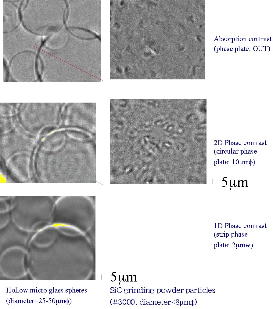
Zernike phase-contrast microscopy was realized using a micro-capillary refractive lens. The high sensitivity of the system was checked by detecting weakly absorbing materials such as the SiC particles with the diameter of less than 5 micrometer.
[ Y. Kohmura, A. Takeuchi, H. Takano, Y. Suzuki and T. Ishikawa, Journal of Phyique. IV France 104, 603-606 (2003), Fig. 1, 4, 5,
©2003 European Physical Society ]
Source of the figure
Original paper/Journal article
Journal title
Y.Kohmura et al., Rev. Sci. Instrum. 70, 4164 (1999)
Figure No.
1,4,5
Technique
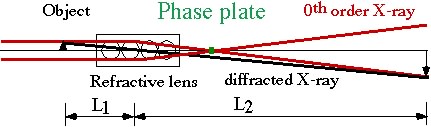
Using a gold quarter-wavelength phase-plate, positive phase contrast images were successfully taken. The microscope system was designed so that the 1D & 2D phase contrast images can be taken by inserting the phase plate and the absorption contrast images without the phase plate.
Source of the figure
No figure
Required time for experimental setup
3 shift(s)
Instruments
| Instrument | Purpose | Performance |
|---|---|---|
| Kouzu Diffractometer | Aligning sample, refractive lens, phase plate |
References
| Document name |
|---|
| Y.Kohmura et al., Rev. Sci. Instrum. 70, 4164 (1999) |
| Y.Kohmura et al., J. Phys. IV France, 104, 603 (2003) |
Related experimental techniques
X-ray Microscopy
Questionnaire
The measurement was possible only in SPring-8. Impossible or very difficult in other facilities.
This solution is an application of a main instrument of the beamline.
Ease of measurement
Middle
Ease of analysis
Easy
How many shifts were needed for taking whole data in the figure?
Four-nine shifts