軟X線照射中に生成するDNA塩基の短寿命ラジカルの観測

Inquiry number
SOL-0000001484
Beamline
BL23SU (JAEA Actinide Science II)
Scientific keywords
| A. Sample category | organic material, atom, molecule, radical, biology, medicine |
|---|---|
| B. Sample category (detail) | organic material, macromolecule, radical, biology (in vitro), biological material |
| C. Technique | absorption and its secondary process |
| D. Technique (detail) | |
| E. Particular condition | polarization (circular), surface, room temperature, low-T (~ liquid N2) |
| F. Photon energy | soft X-ray |
| G. Target information | chemical state, spin/magnetism, photochemical reaction |
Industrial keywords
| level 1---Application area | environment, Pharmaceuticals |
|---|---|
| level 2---Target | drug design, process analytical technology (PAT) |
| level 3---Target (detail) | protein, active pharmaceutical ingredients (API), tablet |
| level 4---Obtainable information | chemical state |
| level 5---Technique |
Classification
M40.40 soft x-ray spectroscopy
Body text
軟X線照射中に生成するラジカルの測定は内殻励起によって生成した短寿命ラジカルを調べることのできるユニークな手法です。この手法を用いることで、構成元素の 内殻励起に特徴的な短寿命のラジカルを測定することができます。世界最高水準の高分解能軟X線分光器は、生体分子の内殻準位を任意に選びながら、生じるラジカルをin situで測定することが可能になります。図に示すのは、グアニンについて測定した酸素K殻励起中に生成した短寿命ラジカルスペクトルです。この結果から、軟X線照射中のみに生成する短寿命ラジカルが存在することが確認され、照射後に分子内に固定される長寿命ラジカルとは異なった由来を持つラジカルであることがわかりました。
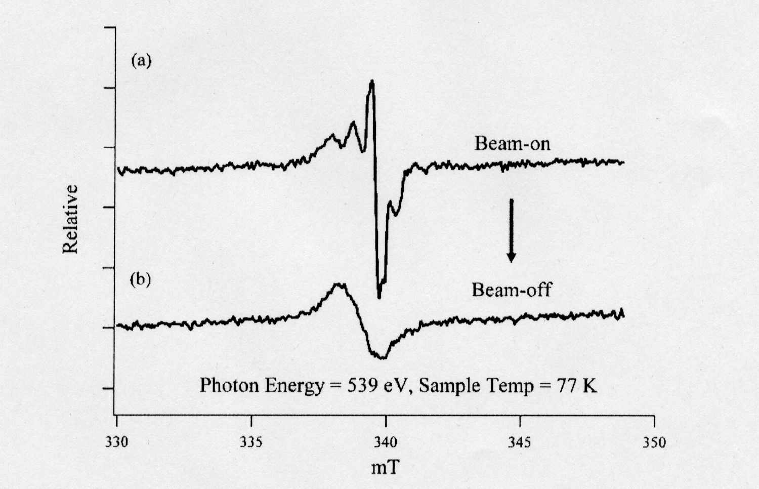
Fig. 1 EPR spectra of guanine during irradiation of soft X-rays (539 eV) at 77K (a) and just after exposure (b). The microwave power was 200W and the width of the magnetic field modulation of 100kHz was 0.5mT.
[ A. Yokoya, K. Akamatsu and K. Fujii, Nuclear Instruments and Methods in Physics Research B 199, 366-369 (2003), Fig. 1,
©2003 Elsevier Science Publisher ]
Source of the figure
Original paper/Journal article
Journal title
A. Yokoya et. al., Nuclear Instruments and Methods in Physics Research B 199 (2003) 366-369
Figure No.
1
Technique
電子常磁性共鳴測定は、ラジカル生成を調べられる簡便な測定方法です。この方法は、固体試料や粉末試料に適応でき、ラジカルの種類や生成量に関する情報を得ることができます。
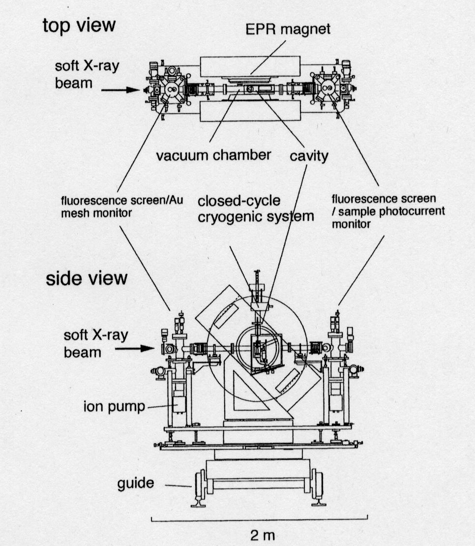
Fig. 2 Illustration of SLEEPRS. The vacuum chamber, in which the microwave cavity is installed, is mounted at the center of the magnet gap (see top view). THe chamber is connected to the beamline vacuum pipe at both the fromt and the rear.
[ A. Yokoya and K. Akamatsu, Nuclear Instruments and Methods in Physics Research A 467-468, 1333-1337 (2001), Fig. 1,
©2001 Elsevier Science Publisher ]
Source of the figure
Original paper/Journal article
Journal title
A. Yokoya et. al., Nuclear Instruments and Methods in Physics Research A 467-468 (2001) 1333-1337
Figure No.
1
Required time for experimental setup
1 hour(s)
Instruments
| Instrument | Purpose | Performance |
|---|---|---|
| 生物物理学的分光装置 | ラジカル生成のその場観測 | X-band |
References
| Document name |
|---|
| A. Yokoya et. al., Nulear Instruments and Methods in Physics Research B 199 (2003) 366-369 |
| A. Yokoya et. al., Nuclear Instruments and Methods in Physics Research A 467-468 (2001) 1333-1337 |
Related experimental techniques
Questionnaire
The measurement was possible only in SPring-8. Impossible or very difficult in other facilities.
Ease of measurement
With a great skill
Ease of analysis
Middle
How many shifts were needed for taking whole data in the figure?
Two-three shifts