Stereograph of the atomic arrangement of graphite

Inquiry number
SOL-0000000904
Beamline
BL25SU (Soft X-ray Spectroscopy of Solid)
Scientific keywords
| A. Sample category | inorganic material, research on method, instrumentation |
|---|---|
| B. Sample category (detail) | metal, alloy, semiconductor, solid-state crystal |
| C. Technique | X-ray diffraction, X-ray elastic scattering, photoemission, photoionization |
| D. Technique (detail) | photoelectron spectra, photoelectron holography |
| E. Particular condition | polarization (circular), 2D imaging, ultra-high vacuum, surface, interface |
| F. Photon energy | soft X-ray |
| G. Target information | local structure, structure analysis, crystal structure, phase transition |
Industrial keywords
| level 1---Application area | Semiconductor |
|---|---|
| level 2---Target | silicon semiconductor, compound semiconductor, catalysis |
| level 3---Target (detail) | |
| level 4---Obtainable information | d-spacing (lattice parameter), interatomic distance, crystal structure, local structure, adsorption |
| level 5---Technique | XPS, diffraction |
Classification
A30.20 surface・interface, A40.40 surface・interface chemistry, A80.12 semiconductor, A80.20 metal ・material, M10.10 single crystal diffraction, M10.30 surface・interface diffraction, M50.10 photoelectron spectroscopy
Body text
Stereoscopic microscopy of atomic arrangement is a unique technique to study the local atomic structure of surface, or impurity. Using this technique, one can measure the local atomic structure of the selected element. The long range order of the sample is not always required. In addition, the technique may have application to light element. The figure shows the stereograph of atomic arrangement of graphite single-crystal. These data reveal the fact that the stereoscopic microscopy can measure the atomic structure with high accuracy.
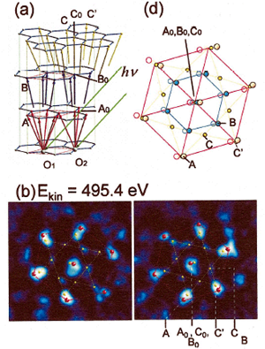
[ F. Matsui, H. Daimon, F. Z. Guo and T. Matsushita, Applied Physics Letters 85, 3737-3739 (2004), Fig. 2,
©2004 American Institute of Physics ]
Source of the figure
Original paper/Journal article
Journal title
Appl. Phys. Lett. 85,3737 (2004)
Figure No.
2
Technique
Stereoscopic microscopy of atomic arrangement is a unique technique to study local atomic structure. Two photoelectron diffraction patterns excited by right and left polarized light form a stereograph of local atomic structure. The technique is applicable to crystal, adsorbates on surface, atom in interface, or impurities in crystal and provides knowledge about the local atomic structure.
Source of the figure
No figure
Required time for experimental setup
1 day(s)
Instruments
| Instrument | Purpose | Performance |
|---|---|---|
| Two-dimensional Display-type Angle-resolved Photoelectron Analyzer (2D-PES) | Stereoscopic microscopy of Atomic arrangement | Energy range 200-1000eV, Energy reslution 0.25eV, Acceptance angle 120deg, Angular resolution 0.6deg |
References
| Document name |
|---|
| Phys. Rev. Lett. 86, 2034 (2001) |
Related experimental techniques
Photoemission, Photoelectron diffraction, Photoelectron holography, Fluorescent X-ray holograply, X-ray diffraction.
Questionnaire
The measurement was possible only in SPring-8. Impossible or very difficult in other facilities.
Ease of measurement
Middle
Ease of analysis
Middle
How many shifts were needed for taking whole data in the figure?
Two-three shifts