XMCD of Ce compounds

Inquiry number
SOL-0000001117
Beamline
BL25SU (Soft X-ray Spectroscopy of Solid)
Scientific keywords
| A. Sample category | inorganic material |
|---|---|
| B. Sample category (detail) | metal, alloy, magnetic material |
| C. Technique | absorption and its secondary process |
| D. Technique (detail) | MCD, LD |
| E. Particular condition | polarization (circular), ultra-high vacuum, low-T (~ liquid He), magnetic field (< 2 T) |
| F. Photon energy | soft X-ray |
| G. Target information | electronic state, spin/magnetism |
Industrial keywords
| level 1---Application area | storage device, industrial material |
|---|---|
| level 2---Target | HD,MO |
| level 3---Target (detail) | magnetic layer |
| level 4---Obtainable information | electronic state, magnetic moment |
| level 5---Technique | XMCD |
Classification
A80.14 magnetic materials, M40.30 XMCD, M40.40 soft x-ray spectroscopy
Body text
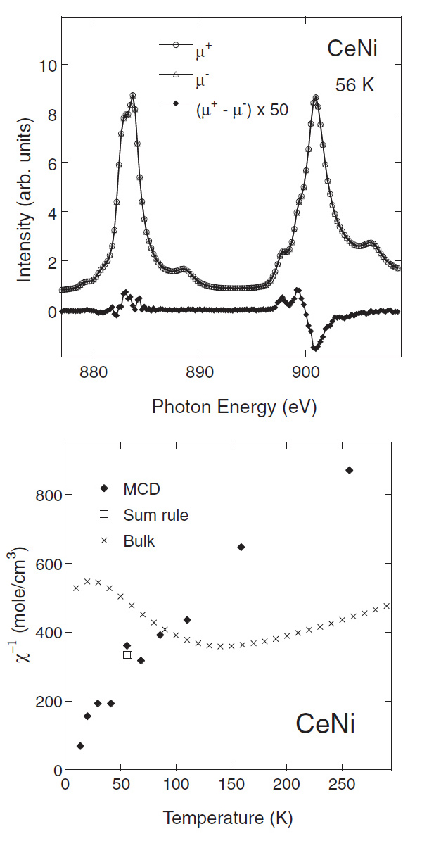
X-ray magnetic circular dichroism (XMCD) is a powerful tool to study magnetic properties of materials. Magnetism of the 4f electrons of Lanthanoid (La - Yb) compounds is favorably investigated by XMCD in soft x-ray region at BL25SU. The element specificity is an great advantage of XMCD, which enables us to obtain magnetic information of target element even when a sample contain several kinds of elements. Upper figure below shows absorption and the XMCD spectra of Ce M4,5-edges in CeNi alloy. Under figure shows inverse of the Ce 4f magnetic susceptibility -1(T ) obtained by XMCD in a magnetic field of H = 1.4 T and the inverse conventional bulk magnetic susceptibility -1(T ). XMCD spectrum is measured clearly in spite of weak magnetization of paramagnetic CeNi. A large discrepancy of -1(T ) behaviors is observed between XMCD and the bulk measurement. It is attributed that the 5d electrons contributes to bulk magnetization in addition to the 4f magnetism detected by XMCD at the Ce M4,5-edges.
[ H. Shiozawa, T. Miyahara, K. Obu, Y. Takayama, H. Ishii, T. D. Matsuda, H. Sugawara, H. Sato, T. Muro, Y. Saitoh, Journal of the Physical Society of Japan 72, 2079-2084 (2003), Fig. 1, 5,
©2003 The Japan Society of Applied Physics ]
Source of the figure
Original paper/Journal article
Journal title
Shiozawa et al., J. Phys. Soc. Jpn. 72 (2003), 2079-2085.
Figure No.
1,5
Technique
XMCD technique is applied to the present study. Magnetic field of 1.4 T was applied to a CeNi sample so that effective XMCD signal can be detected. More advanced XMCD measurements is possible using the 4th. experimental station (electromagnet type XMCD spectrometer) at BL25SU, though the previous XMCD spectrometer using permanent magnet of 1.4 T was used for the present experiments.
Source of the figure
No figure
Required time for experimental setup
6 hour(s)
Instruments
| Instrument | Purpose | Performance |
|---|---|---|
| Magnetic Circular Dichroism Measurement System (MCD) | Measurements of XMCD | 1.4 T (fixed), 40K - 300K |
References
Related experimental techniques
ESMH using hard x-ray at the BL39XU, VSM, SQUID, PEEM, Kerr effect, Mossbauer spectroscopy
Questionnaire
This solution is an application of a main instrument of the beamline.
Similar experiments account for more than 30% of the beamline's subject.
Ease of measurement
Easy
Ease of analysis
Middle
How many shifts were needed for taking whole data in the figure?
Two-three shifts