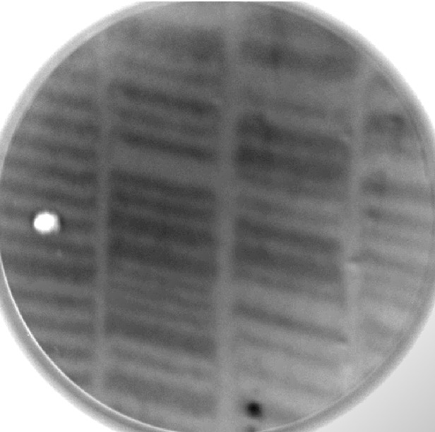
PEEM image of recorded magnetic domain in HDD

問い合わせ番号
SOL-0000001164
ビームライン
BL25SU(軟X線固体分光)
学術利用キーワード
| A. 試料 | 無機材料, 計測法、装置に関する研究 |
|---|---|
| B. 試料詳細 | 金属・合金, 磁性体, 膜 |
| C. 手法 | 吸収、及びその二次過程 |
| D. 手法の詳細 | MCD, LD, 光電子顕微鏡 |
| E. 付加的測定条件 | 偏光(円、楕円), 高分解能画像計測(顕微鏡), 超高真空, 表面, 室温 |
| F. エネルギー領域 | 軟X線(<2 keV) |
| G. 目的・欲しい情報 | スピン・磁性構造 |
産業利用キーワード
| 階層1 | 記憶装置 |
|---|---|
| 階層2 | HD、MO |
| 階層3 | 磁性層 |
| 階層4 | 磁化 |
| 階層5 | XMCD, 磁気散乱, 磁気コンプトン散乱, PEEM, PEEM, イメージング |
分類
A30.20 表面界面物性, A80.14 磁性材料, A80.30 無機材料, M40.30 磁気吸収, M40.40 軟X線分光, M50.20 光電子顕微鏡(PEEM)
利用事例本文
Photoemission electron microscope (PEEM) is a new technique providing microscopic images of contrast of secondary electrons intensity emitted by photo-irradiation.Element distribution, chemical state of microscopic area, and element specific magnetic domain, so on, are obtained by means of PEEM with synchrotron x-rays. PEEM has advantages of the real time image acquisition by means of CCD camera and less sample damages comparing to SEM.
Figure shows a contrast of magnetic circular dichroism (MCD) at the Co L3-edge of a recording disk installed inside HDD.The 3.5 inch HDD has 250MB storage in total and the 10 micrometer track width. This is a demonstrative image of element specific magnetic domain by means of PEEM.
画像ファイルの出典
私信等、その他
詳細
2005 Summer School at SPring-8
測定手法
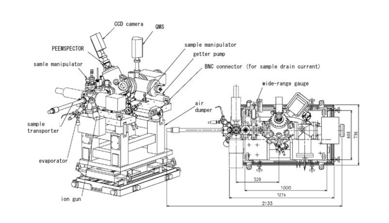
The PEEMSPECTOR installed at BL25SU was used. The element specific magnetic domain image at the Co L3-edge was obtained as a difference between two PEEM images recorded using the left- and right-handed soft x-rays.
画像ファイルの出典
BL評価レポート
ページ
56
測定準備に必要なおおよその時間
12 時間
測定装置
| 装置名 | 目的 | 性能 |
|---|---|---|
| Photoemission Electron Microscope (PEEM) | Recording element specific magnetic domain | 35 - 100nm resolution |
参考文献
関連する手法
Scaning electron microscope (SEM), Magnetic force microscope (MFM), Kerr microscope, X-ray Magnetic circular dichroism (XMCD)
アンケート
最近2年以内に導入した装置を使った事例
測定の難易度
初心者でもOK
データ解析の難易度
初心者でもOK
図に示した全てのデータを取るのにかかったシフト数
2~3シフト