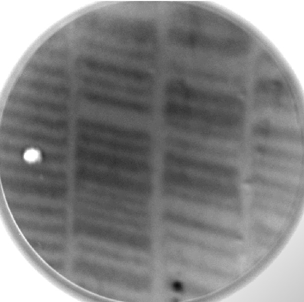
PEEM image of recorded magnetic domain in HDD

Inquiry number
SOL-0000001164
Beamline
BL25SU (Soft X-ray Spectroscopy of Solid)
Scientific keywords
| A. Sample category | inorganic material, research on method, instrumentation |
|---|---|
| B. Sample category (detail) | metal, alloy, magnetic material, membrane |
| C. Technique | absorption and its secondary process |
| D. Technique (detail) | MCD, LD, PEEM |
| E. Particular condition | polarization (circular), X-ray microscopy, ultra-high vacuum, surface, room temperature |
| F. Photon energy | soft X-ray |
| G. Target information | spin/magnetism |
Industrial keywords
| level 1---Application area | storage device |
|---|---|
| level 2---Target | HD,MO |
| level 3---Target (detail) | magnetic layer |
| level 4---Obtainable information | magnetic moment |
| level 5---Technique | XMCD, magnetic scattering, magnetic Compton scattering, PEEM, PEEM, imaging |
Classification
A30.20 surface・interface, A80.14 magnetic materials, A80.30 inorganic material, M40.30 XMCD, M40.40 soft x-ray spectroscopy, M50.20 PEEM
Body text
Photoemission electron microscope (PEEM) is a new technique providing microscopic images of contrast of secondary electrons intensity emitted by photo-irradiation.Element distribution, chemical state of microscopic area, and element specific magnetic domain, so on, are obtained by means of PEEM with synchrotron x-rays. PEEM has advantages of the real time image acquisition by means of CCD camera and less sample damages comparing to SEM.
Figure shows a contrast of magnetic circular dichroism (MCD) at the Co L3-edge of a recording disk installed inside HDD.The 3.5 inch HDD has 250MB storage in total and the 10 micrometer track width. This is a demonstrative image of element specific magnetic domain by means of PEEM.
Source of the figure
Private communication/others
Description
2005 Summer School at SPring-8
Technique
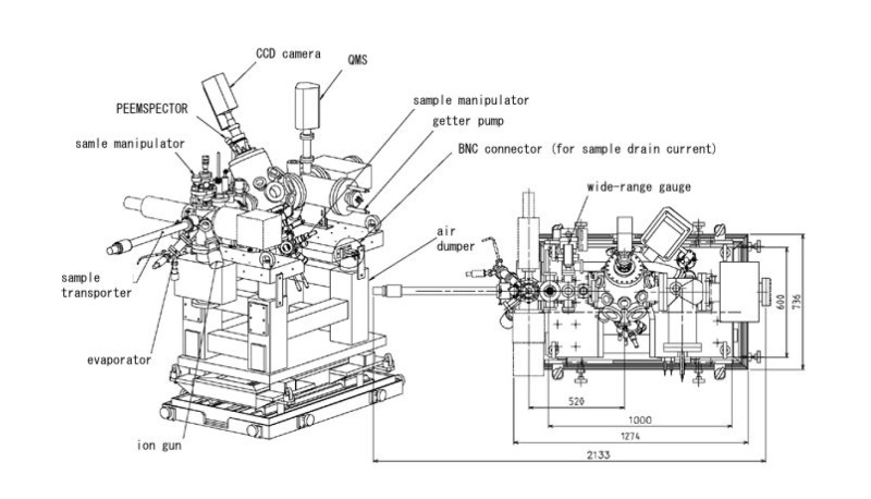
The PEEMSPECTOR installed at BL25SU was used. The element specific magnetic domain image at the Co L3-edge was obtained as a difference between two PEEM images recorded using the left- and right-handed soft x-rays.
Source of the figure
Beamline Report
Page
56
Required time for experimental setup
12 hour(s)
Instruments
| Instrument | Purpose | Performance |
|---|---|---|
| Photoemission Electron Microscope (PEEM) | Recording element specific magnetic domain | 35 - 100nm resolution |
References
Related experimental techniques
Scaning electron microscope (SEM), Magnetic force microscope (MFM), Kerr microscope, X-ray Magnetic circular dichroism (XMCD)
Questionnaire
This solution is application of a new instrument installed in the past two years.
Ease of measurement
Easy
Ease of analysis
Easy
How many shifts were needed for taking whole data in the figure?
Two-three shifts