摘出拍動ラット心臓での冠状動脈の応答測定

Inquiry number
SOL-0000001529
Beamline
BL28B2 (White Beam X-ray Diffraction)
Scientific keywords
| A. Sample category | biology, medicine |
|---|---|
| B. Sample category (detail) | biology (in vitro) |
| C. Technique | absorption and its secondary process |
| D. Technique (detail) | |
| E. Particular condition | 2D imaging, time-resolved (ms) |
| F. Photon energy | X-ray (4-40 keV) |
| G. Target information |
Industrial keywords
| level 1---Application area | Pharmaceuticals |
|---|---|
| level 2---Target | drug design |
| level 3---Target (detail) | drug |
| level 4---Obtainable information | molphology |
| level 5---Technique | imaging |
Classification
A80.90 others
Body text
心臓冠状動脈は拍動による動きのため、特に心拍数が高い小動物について、高精度な計測が難しくなっています。しかし、放射光の利用によって、短時間 (2 msec) での撮像が可能となり、ラット心臓でその動きを止めた高精度計測が実現しました。これにより血管作用薬での冠状動脈の拡張・収縮反応を高精度で計測可能となりました。
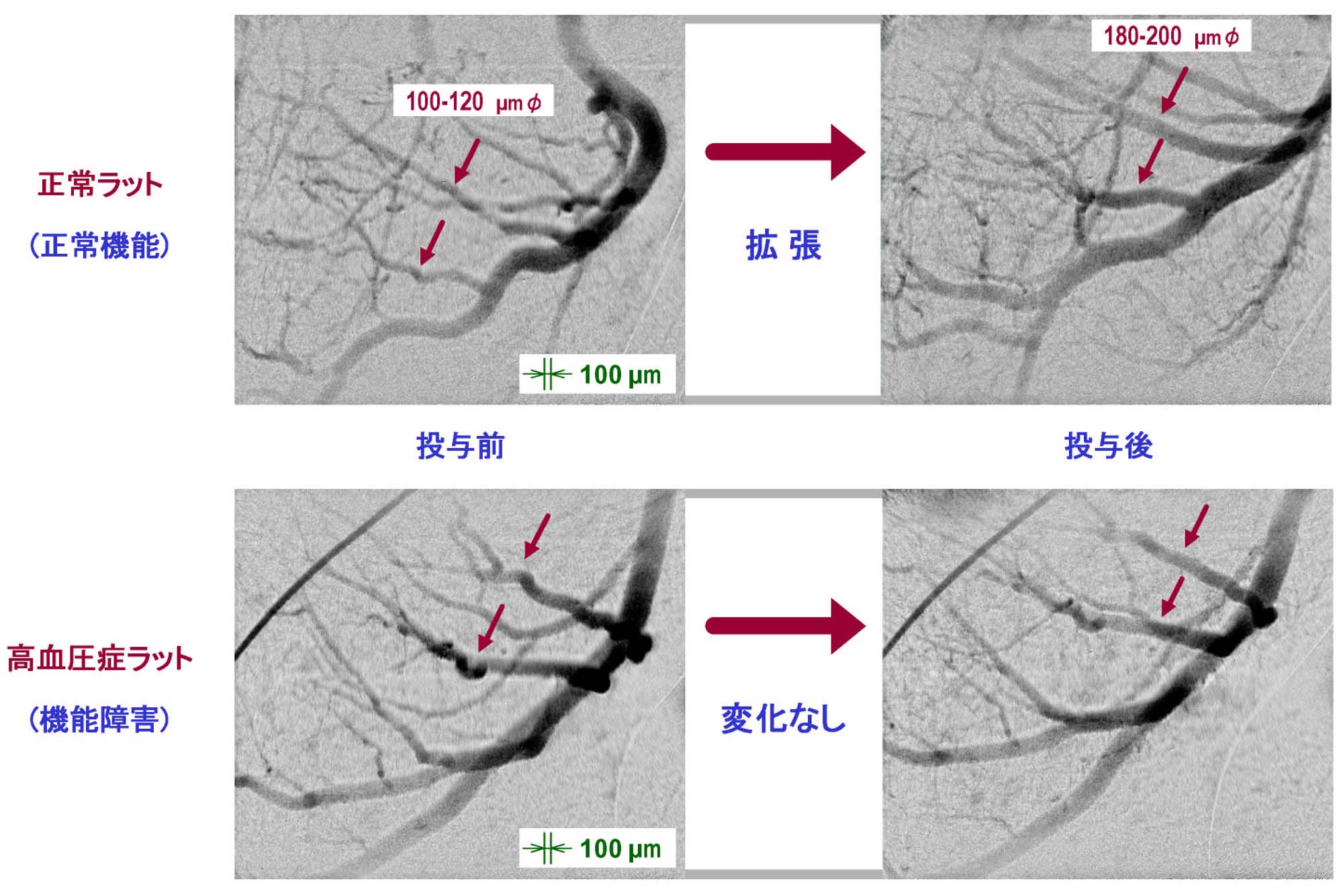
図1 血管作用薬アセチルコリン投与前後での冠動脈径の変化
[ K. Umetani, K. Fukushima and K. Sugimura, 映像情報メディア学会誌 58, 344-351 (2004), Fig. 8,
©2004 映像情報メディア学会 ]
Source of the figure
Original paper/Journal article
Journal title
映像情報メディア学会誌 58(3) 344-351 (2004)
Figure No.
8
Technique
BL28B2では一結晶分光器で単色化した高強度の単色X線を利用できるため、動物実験においては心臓血管のパルスX線を使った実時間観察も可能となっています。このため、X線直接変換型サチコン撮像管を使った撮影視野が4.5 mm×4.5 mmで空間解像度が6 µmの画像検出器を開発し、さらに回転円盤型X線シャッターを新たに開発してパルスX線撮影を可能としました。これを使い血管内に造影剤を注入して血管を画像化する血管造影で、ラット心臓において心筋に血液を供給する冠状動脈の撮影を行います。
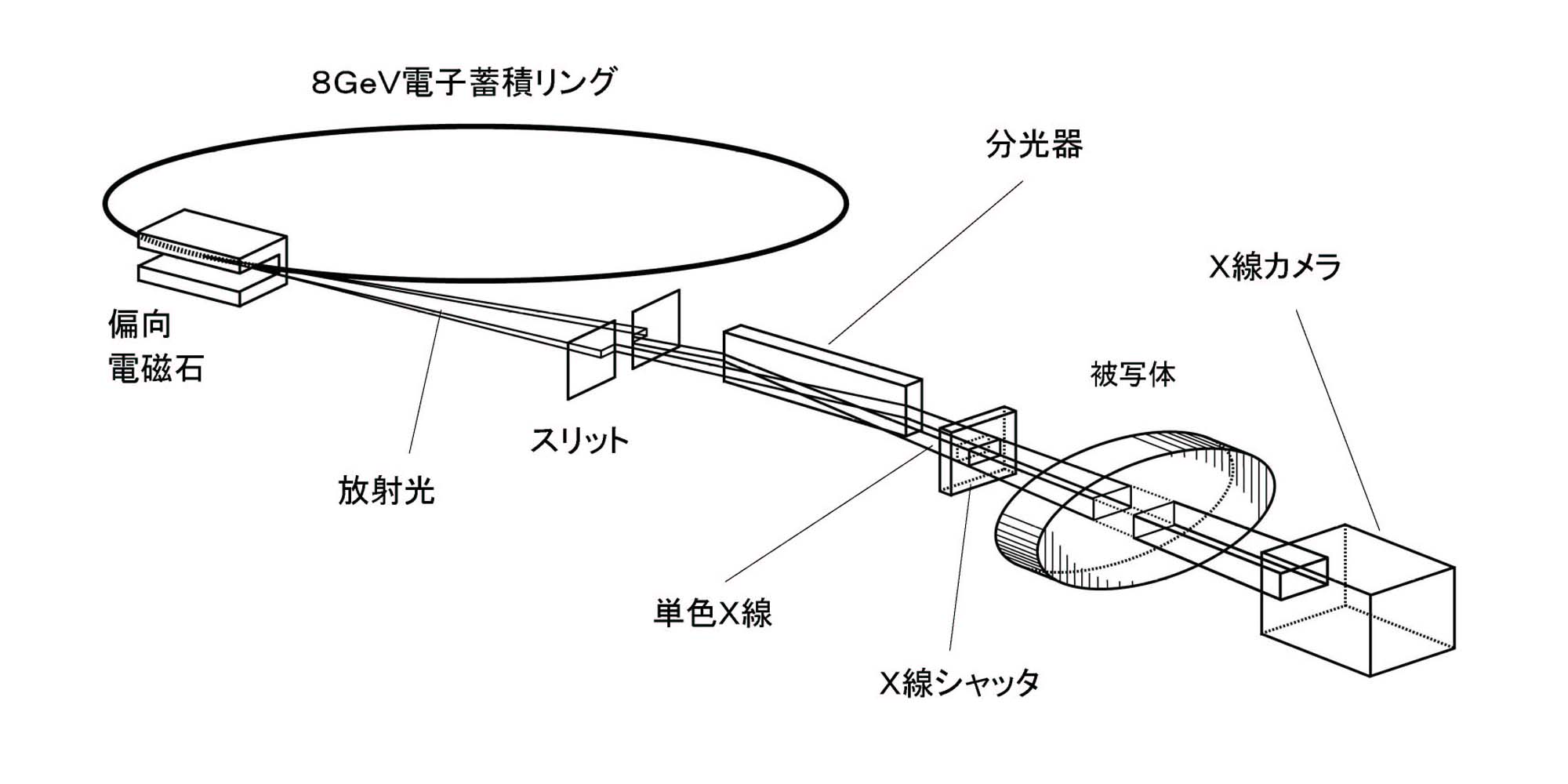
図2 血管造影での装置構成
Source of the figure
Presentation material for Beamline Report
Required time for experimental setup
3 hour(s)
Instruments
| Instrument | Purpose | Performance |
|---|---|---|
| 血管造影装置 | 微小血管造影 | 解像度6~10μm |
References
| Document name |
|---|
| Journal of Institute of Image Information and Television Engineers 58(3) 344-351 (2004). |
Related experimental techniques
特になし
Questionnaire
The measurement was possible only in SPring-8. Impossible or very difficult in other facilities.
Ease of measurement
Middle
Ease of analysis
Middle
How many shifts were needed for taking whole data in the figure?
Two-three shifts