Scanning X-ray fluorescence microscopy
Inquiry number
SOL-0000000975
Beamline
BL29XU (RIKEN Coherent X-ray Optics)
Scientific keywords
| A. Sample category | inorganic material, organic material, biology, medicine |
|---|---|
| B. Sample category (detail) | metal, alloy, semiconductor, insulator, ceramics, organism, cell, biological material |
| C. Technique | fluorescent X-rays |
| D. Technique (detail) | trace-element |
| E. Particular condition | microbeam (1-10 µm), microbeam (sub-µm), 2D imaging |
| F. Photon energy | X-ray (4-40 keV) |
| G. Target information | structure analysis, function and structure |
Industrial keywords
| level 1---Application area | environment, Pharmaceuticals |
|---|---|
| level 2---Target | drug design, Environmental material |
| level 3---Target (detail) | organism |
| level 4---Obtainable information | element distribution |
| level 5---Technique | imaging |
Classification
A80.20 metal ・material, A80.30 inorganic material, A80.32 organic material, A80.50 Pharmaceuticals, A80.90 others
Body text
Scanning x-ray fluorescence microscopy is an efficient technique to study element distribution. Element mapping with a spatial resolution of tens of nm can be performed with a Kirkpatrick-Baez type x-ray focusing system composed of ultra-precise x-ray total reflection mirrors.
The following figure shows the element maps of CDDP-sensitive and -resistant cancer cells. A 15 keV x-ray beam was focused to a spot size of 1.5 m (H) 0.75 m (W) on the sample. These data helped to propose a novel cancer treatment.
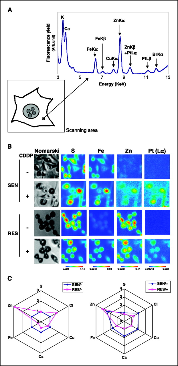
Fig. Element maps of cancer cells by scanning x-ray fluorescence microscopy
[ M. Shimura, A. Saito, S. Matsuyama, T. Sakuma, Y. Terui, K. Ueno, H. Yumoto, K. Yamamura, H. Mimura, Y. Sano, M. Yabashi, K. Tamasaku, K. Nishio, Y. Nishino, K. Endo, K. Hatake, Y. Mori and Y. Ishizuka, Cancer Research 65, 4998-5002 (2005), Fig. 1,
©2005 American Society of Cancer Research ]
Source of the figure
Original paper/Journal article
Journal title
M. Shimura, A. Saito, S. Matsuyama, T. Sakuma, Y. Terui, K. Ueno, H. Yumoto, K. Yamauchi, K. Yamamura, H. Mimura, Y. Sano, M. Yabashi, K. Tamasaku, K. Nishio, Y. Nishino, K. Endo, K. Hatake, Y. Mori, Y. Ishizaka, and T. Ishikawa, Cancer Res. 65, 4998-5002 (2005)
Figure No.
1
Technique
Two-dimensional focusing of an x-ray beam is performed using two focusing mirrors in the Kirkpatrick-Baez configuration. Fluorescent X-ray spectroscopy is performed using a silicon drift detector.
Source of the figure
No figure
Required time for experimental setup
hour(s)
Instruments
| Instrument | Purpose | Performance |
|---|---|---|
| Kirkpatrick-Baez type x-ray focusing system | ||
| silicon drift detector |
References
| Document name |
|---|
| M. Shimura, A. Saito, S. Matsuyama, T. Sakuma, Y. Terui, K. Ueno, H. Yumoto, K. Yamauchi, K. Yamamura, H. Mimura, Y. Sano, M. Yabashi, K. Tamasaku, K. Nishio, Y. Nishino, K. Endo, K. Hatake, Y. Mori, Y. Ishizaka, and T. Ishikawa, Cancer Res. 65, 4998-5002 (2005) |
Related experimental techniques
Questionnaire
With user's own instruments.
Ease of measurement
With a great skill
Ease of analysis
With a great skill
How many shifts were needed for taking whole data in the figure?
Four-nine shifts