Scanning X-ray magnetic microprobe


Inquiry number
SOL-0000001036
Beamline
BL39XU (X-ray Absorption and Emission Spectroscopy)
Scientific keywords
| A. Sample category | research on method, instrumentation |
|---|---|
| B. Sample category (detail) | magnetic material |
| C. Technique | absorption and its secondary process |
| D. Technique (detail) | XAFS, XANES, MCD, LD |
| E. Particular condition | polarization (circular), microbeam (1-10 µm), 2D imaging, magnetic field (< 2 T), room temperature |
| F. Photon energy | X-ray (4-40 keV) |
| G. Target information | spin/magnetism |
Industrial keywords
| level 1---Application area | storage device |
|---|---|
| level 2---Target | HD,MO |
| level 3---Target (detail) | magnetic layer, magnetic head, spin valve |
| level 4---Obtainable information | magnetic moment, magnetic anisotropy, interface magnetic structure |
| level 5---Technique | XAFS, XMCD, imaging |
Classification
A80.14 magnetic materials, M40.30 XMCD
Body text
Scanning X-ray magnetic microprobe is a unique tool to obtain a two-dimensional magnetic image of specific magnetic elements comprised in the sample. Available spatial resolution is approximately 2 m. The technique is applicable to magnetic materials containing 3d transition metal, rare earth, and 5d noble metal elements. It also allows X-ray magnetic circular dichroism (XMCD) or XAFS measurements for a specific area of a few m square. The figure shows a magnetic image taken for a CoCrPtB in-plane magnetization film. Magnetic recording patterns were observed as an XMCD contrast of X-ray fluorescence intensity at the Pt L3 edge. The stripe of 2.4 m width was clearly resolved.
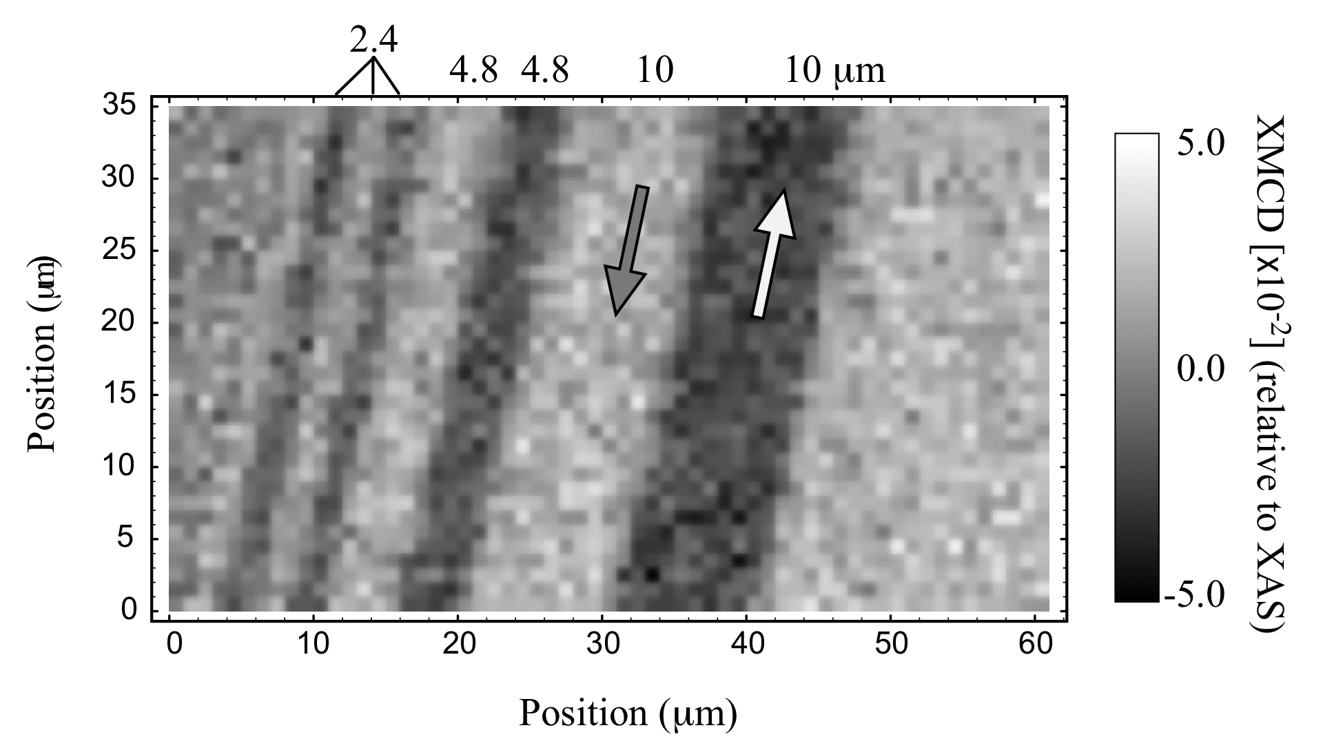
Fig. Magnetic image of a CoCrPtB in-plane magnetization film,
taken using the scanning X-ray magnetic microprobe.
Source of the figure
Bulletin from SPring-8
Bulletin title
ナノテクノロジー総合支援プロジェクト研究成果報告書 Vol.4, 2004年A
Page
p. 147
Technique
The scanning X-ray magnetic microprobe consists of a diamond X-ray phase plate and X-ray focusing mirrors (KB mirror). Monochromatic X-rays from the beamline monochromator are converted into circular polarization by the phase plate, and then focused onto a sample by the KB mirror. The focused spot size is 2 m. X-ray magnetic circular dichroism (XMCD) signal is recorded by monitoring X-ray fluorescence intensity from the sample with changing the incident X-ray photon helicities; the XMCD signal reflects the magnitude or direction of sample magnetization. A magnetic image is taken by two-dimensionally scanning the sample position using high-precision motorized stages.
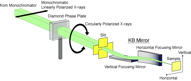
Fig. A schematic of the scanning X-ray magnetic microprobe.
Source of the figure
Bulletin from SPring-8
Bulletin title
ナノテクノロジー総合支援プロジェクト研究成果報告書 Vol. 4, 2004年A
Page
p. 146
Required time for experimental setup
3 shift(s)
Instruments
| Instrument | Purpose | Performance |
|---|---|---|
| Scanning X-ray magnetic microprobe | Measurement of 2D magnetic image, XMCD spectroscopy in a minute sample area | Focused X-ray beam spot 2 microns, photon flux 10^10 photons/s |
References
| Document name |
|---|
| M. Takagaki, M. Suzuki, N. Kawamura, H. Mimura, and T. Ishikawa, The 8th International Conference on X-ray Microscopy (XRM2005), 26-30 July 2005, Himeji, Japan. |
Related experimental techniques
magnetic force microscope, MFM, photoelectron emission microscope, PEEM, X-ray transmission microscope
Questionnaire
The measurement was possible only in SPring-8. Impossible or very difficult in other facilities.
This solution is an application of a main instrument of the beamline.
This solution is application of a new instrument installed in the past two years.
Ease of measurement
Easy
Ease of analysis
Easy
How many shifts were needed for taking whole data in the figure?
Two-three shifts