Cryogenic X-ray diffraction experiment under ultra-low temperature using helium gas flow cryostream cooler
Inquiry number
SOL-0000001180
Beamline
BL41XU (Macromolecular Crystallography I)
Scientific keywords
| A. Sample category | biology, medicine |
|---|---|
| B. Sample category (detail) | biomolecule, crystal, protein, nucleic acid, pharmaceuticals |
| C. Technique | X-ray diffraction |
| D. Technique (detail) | single crystal |
| E. Particular condition | low-T (~ liquid He) |
| F. Photon energy | X-ray (4-40 keV) |
| G. Target information | molecular structure, local structure, structure analysis, function and structure |
Industrial keywords
| level 1---Application area | environment, Pharmaceuticals |
|---|---|
| level 2---Target | catalysis, drug design, process analytical technology (PAT), food |
| level 3---Target (detail) | protein, drug |
| level 4---Obtainable information | chemical state, supra-molecular assemblies, absolute configuration |
| level 5---Technique | diffraction |
Classification
A80.50 Pharmaceuticals, A80.60 food、daily necessaries, M10.10 single crystal diffraction
Body text
There are large amount of water molecules in a protein crystal, and these are changed to hydroxyl radicals by X-ray exposure. It is thought that X-ray damages are caused by diffusing of those radicals while destroying the tertiary structure of the protein or intermolecular interaction in the crystal. X-ray damage is observed as a decrease in the X-ray diffraction ability, a deterioration of diffraction spots or an increase of mosaic spread. These prevent a precise data collection. Cooling the sample about to 100K is effective in the decrease of X-ray damage to prevent radicals from diffusing.
In the X-ray diffraction experiment using generator in laboratory or bending magnet beamline in past facilities, X-ray damage was hardly observed by cooling samples to 100K. However, X-ray damage was occurred in 100K using beamlines at third-generation synchrotron facilities.
In this case, sufficient diffraction data could not be collected in 100K by X-ray damage. Then, the sample was cooled to 35K by helium gas flow cryostream cooler, and precise diffraction data sets of ArcTGT・tRNAVal complex from P.horikoshii were collected successfully.
By analyzing these data, form of tRNA was newly discovered (Fig. 1).
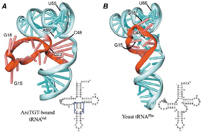
Figure 1. Drawings of newly discovered form (A) and conventional L form (B)of tRNA
[ R. Ishitani, O. Nureki, N. Nameki, N. Okada, S. Nishimura and S. Yokoyama, Cell 113, 383-394 (2003), Fig. 3,
©2003 Cell Press ]
Source of the figure
Original paper/Journal article
Journal title
R. Ishitani, et al., Cell 113, 383-394 (2003)
Figure No.
3
Technique
Source of the figure
No figure
Required time for experimental setup
3 hour(s)
Instruments
| Instrument | Purpose | Performance |
|---|---|---|
| Protein Crystal Diffractometer | To record diffraction data | |
| Helium gas flow cryostream cooler | To cool sample |
References
| Document name |
|---|
| R. Ishitani, et al., Cell 113, 383-394 (2003) |
Related experimental techniques
Questionnaire
This solution is an application of a main instrument of the beamline.
Ease of measurement
Middle
Ease of analysis
Middle
How many shifts were needed for taking whole data in the figure?
Four-nine shifts