The First Successful Four-Dimensional Visualization of Fuel Cell Pt Catalyst’s Distribution and Chemical State (Press Relase)
- Release Date
- 14 Sep, 2012
- BL47XU (HAXPES / uCT)
Institute for Molecular Science (National Institute of Natural Science)
Japan Synchrotron Radiation Research Institute
|
A research group* at Institute of Molecular Science (IMS) and Japan Synchrotron Radiation Research Institute (JASRI) successfully constructed the world’s first four-dimensional visualization of the internal state of a fuel cell membrane and membrane-electrode assembly (MEA), utilizing the facilities provided by SPring-8*1 (one of the world’s most advanced large-scale synchronous radiation facilities). The 4D visualization (three spatial axes and one energy axis) provides intangible information, i.e., the distribution of the catalyst and the chemical state inside the fuel cell membrane and MEA, with a visual perspective. The fuel cell is coming into widespread use both in households (e.g., “Ene Farm” residential fuel cell) and industry (typically, commercialization for automobiles) as a next-generation energy source. Still, it faces challenges, including the upgrade of power generation efficiency and reduction of undesirable effects (e.g. elution/degradation of precious Pt catalyst on the cathode). As the fuel cell MEA is a complex heterogeneous system, the development of a method to probe inside it nondestructively in order to identify spatial locations of Pt catalyst’s leaching out and degradation have been a very challenging task. * IMS: Mizuki Tada (associate professor), Takahiro Saida (post doctorate fellow) The research group used a novel method - X-ray laminography*2 XAFS*3, which is newly developed in SPring-8 - to obtain hitherto intangible information on cathode catalyst layers inside the fuel cell MEA. The result was the first successful visualization of Pt catalyst distribution and chemical state in the cathode catalyst layers in 3D perspectives. The results obtained are expected to shed new light on the degradation mechanism of fuel cell catalyst, and accelerate the development of materials with higher durability for use in fuel cells. The research was conducted as part of a NEDO project (Development of PEFC Technologies for Commercial Promotion / Fundamental Technology Development / Analysis of structure, reaction, and material transport of MEA materials) and the results were published in the on-line version of “Angew. Chem. Int. Ed.” on 11th of September (German time). Publication: |
<<Figures>>
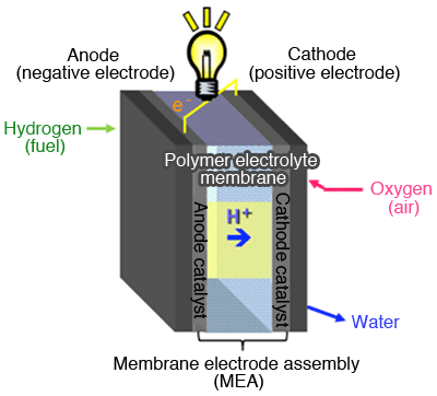
Anode catalyst layer, Polymer electrolyte membrane, MEA (membrane-electrode assembly) is an assembled body of cathode catalyst layers joined together. A catalyst is used both in anode and cathode electrode reactions.

as revealed by X-ray laminography XAFS measurement.
(Upper) 3D plot of the amount of Pt catalyst (Lower) The plot above is projected onto the xy, yz, and zx planes. These clearly indicate the state of degradation on each plane. (Left) MEA before aging procedures. (Right) MEA after 200 cycles of voltage operation. In the MEA, after aging, many spots formed by local coagulation of Pt are observed over the entire area of the cathode catalyst layer.
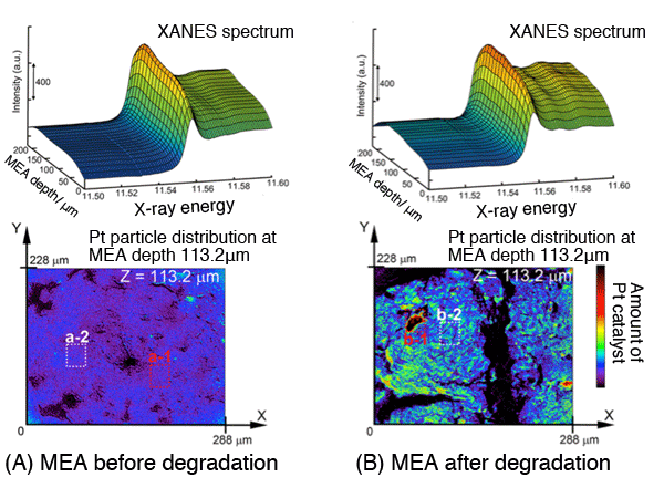
(along the membrane junction) of MEA.(Lower) 2D imaging of Pt catalyst quantity
present on a plane separated from electrolyte membrane by 113.2μm.
MEA after aging (B) shows uneven Pt catalyst distribution, and large coagulations of Pt are observed in places as shown in (b-1) in the right-bottom figure. XANE spectra corresponding to these spots allow oxidation-state analysis of Pt catalyst.
<<Glossary>>
*1 SPring-8
A RIKEN facility located in Harima Science Garden City (Hyogo prefecture) is capable of producing the world's highest intensity synchronous radiation. The management and promotion of utilization of this facility are undertaken by JASRI. The name “SPring-8” comes from “Super Photon ring-8GeV.” An electron flying at nearly the speed of light, if deflected from its original trajectory through the effect exerted by a magnet, emits an electromagnetic wave in a direction tangential to its trajectory, which is called radiation light (or synchrotron radiation). At present, there are three “3rd Generation” large scale synchronous radiation facilities in the world: SPring-8 (Japan), APS (USA) and ESRF (France). The acceleration energy available at SPring-8 (8 billion electron volts) enables the provision of an extremely wide spectrum of radiation light: from far infrared to visible, vacuum ultraviolet, and soft X-ray up to hard X-ray. SPring-8 provides a theater for collaborative works involving researchers inside and outside Japan, and the research conducted at this facility cover such diverse areas as material science, geoscience, life science, environmental science, and various applications in industrial sectors.
*2 X-ray laminography
A type of X-ray CT that performs computer-aided reconstruction of cross-sectional imaging and 3D shapes based on transfer images of the specimen. X-ray imaging techniques normally rotate the specimen to obtain transfer images. One peculiar aspect of this technique includes the fact that it uses X-ray incident angles inclined to, not orthogonal to, the rotation axis. This enables nondestructive measurement of the 3D structure of the region of interest in a plate-like specimen (membrane, PCB, and others) that presents an imaging field’s cross-sectional area typically too large for the conventional X-ray CT method.
*3 XAFS
XAFS is an acronym for X-ray Absorption Fine Structure. X-ray irradiation with a specific range of energy - obtained from synchronous radiation - onto a material gives an X-ray absorption spectrum. Analysis of XANES (X-ray absorption near-edge structure) of this spectrum provides useful information regarding the symmetry and valency of the elements contained in the material. EXAFS (extended X-ray absorption fine structure) analysis also provides valuable information regarding the local configuration of the target element, i.e. how many and what types of atoms are located at what distances from the target element. XAFS is virtually the only technique capable of determining structures that lacks long-range order, typically catalysts. XAFS using hard X-rays enables in-situ structural analysis under actual conditions where catalytic reactions are taking place.
|
For more information, please contact: |

- Previous Article
- An Atomic-Level Analysis of the Mechanism of Proteins Controlling the Iron Concentration inside a Cell (Press Release)
- Current article
- The First Successful Four-Dimensional Visualization of Fuel Cell Pt Catalyst’s Distribution and Chemical State (Press Relase)