Topic 16: Developing High-Resolution Bulk-Sensitive Photoelectron Spectroscopy
Unraveling Anomalous Physical Properties of Strongly-Correlated Electron Materials
Exposing a solid to light causes the electrons in the solid that receive the irradiated light energy to be emitted. This phenomenon is called the photoelectric effect, and the emitted electrons are called photoelectrons. Photoelectron spectroscopy refers to an energy measurement of the emitted photoelectrons to examine the states of electrons in a solid. Dr. Shigemasa Suga,1) (Professor, Osaka University, Japan), a leading authority on photoelectron spectroscopy in Japan, received the Helmholtz-Humboldt Research Award in 2008 for his outstanding long-term contributions in this field. This international prize is awarded to active researchers who have delivered notable academic accomplishments and are expected to continue to actively participate at the forefront of their research field. The research he conducted at SPring-8 played a crucial role in his award.
1) Currently Professor Emeritus, Osaka University.
The Outstanding Achievements of Dr. Suga are Internationally Acknowledged
Dr. Shigemasa Suga, who received the Eugen and Ilse Seibold Prize (Deutsche Forschungsgemeinschaft, Germany) and the Shimadzu Prize (Shimadzu Foundation, Japan), is the first Japanese citizen to be awarded the Helmholtz-Humboldt Research Award. The Helmholtz-Humboldt Research Award is an international honor funded by the Helmholtz Association and Alexander von Humboldt Foundation (Germany).
“The major research topic of the award is research on solid-state physics using bulk-sensitive photoelectron spectroscopy. Bulk refers to the inside of solid state materials,” explains Dr. Suga. Using photoelectron spectroscopy, he was the first to successfully reveal the bulk electronic states of strongly-correlated electron materials. (These materials have strong interactions between electrons.) In 1976, Dr. Suga began researching photoelectron spectroscopy using synchrotron radiation when he was an Associate Professor at the Institute for Solid State Physics, the University of Tokyo, Japan. In 1989, he was appointed a Professor at Osaka University and served as a Visiting Professor at the High Energy Physics Laboratory (KEK)2), Tsukuba, Japan. Throughout his career, Dr. Suga contributed to the development of synchrotron radiation facilities and was an advocate of synchrotron radiation research in Japan. He was actively involved with the entire construction process of SPring-8 from planning to launching the spectroscopic and experimental systems. His goal was that Japan becomes a global leader in bulk-sensitive photoelectron spectroscopy using soft X-rays as a light source. The term “soft” refers to X-rays with lower energies (< 2 keV: keV = 103 eV)3) or longer wavelengths (> 0.6 nm: nm = 10-9 m) compared to common X-rays.
Since the mid-1990s, Dr. Suga along with young researchers and graduate students that he mentored at The University of Tokyo and Osaka University have fully dedicated themselves to developing bulk-sensitive photoelectron spectroscopy. In particular, SPring-8 took the initiative for soft X-ray spectroscopy by utilizing twin-helical undulators. Twin-helical undulators are devices that produce circular polarized synchrotron radiation by helically heaving electron beams with the use of the surrounding magnetic field. Since their inception, the Soft X-ray Spectroscopy of Solid Beamline (BL25SU) at SPring-8 and a high-resolution photoelectron spectroscopic detector (Fig. 1) installed in this beamline have been maintained as the world’s best performing soft X-ray spectroscopy facility. As anticipated, these endeavors at SPring-8 have resulted in successfully developing bulk-sensitive photoelectron spectroscopy as a new field.
2) Currently the High Energy Accelerator Research Organization (KEK).
3) eV = electron volt.
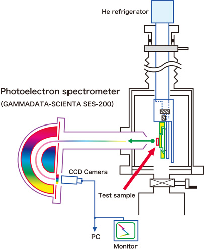
This detector is comprised of a measurement tank and a photoelectron spectrometer. Measurement tank holds the test sample while it is irradiated with synchrotron radiation (soft X-rays), and the spectrometer selectively measures the kinetic energy and intensity of the photoelectrons. Photoelectrons emitted from test samples pass through a long cylindrical-shaped electron lens (high-energy photoelectrons are decelerated) and enter a hemispherical fixture with a static electric field. When a low voltage is applied on the outside and high voltage is applied on the inside of this cup-shaped hemispherical fixture, a static electric field is produced in the hollow portion, bending the trajectory of the entering photoelectrons. By adjusting the voltage, only photoelectrons with specific energy can pass through a hollow portion and be detected (and counted) by a photoelectron detector (CCD camera) placed at the exit.
Successfully Measuring the Electronic States in a Solid
Although commonly used existing spectrometers can observe the surface of a solid using low-energy light, they cannot achieve a sufficient high-resolution measurement using high-energy light. Thus, little useful information has been obtained about the bulk electronic states (electrons inside a material). However at the end of the 20th century, Dr. Suga, Dr. Akira Sekiyama4) (Assistant Professor, Osaka University), and colleagues were the first to reveal the real bulk electronic states of two types of solids (CeRu2Si2 and CeRu2) using the ultra-high resolution soft X-ray monochromator and the high-resolution photoelectron spectrometer that they designed and developed (Fig. 2).
The technique, which Dr. Suga and colleagues developed by bringing all relevant Japanese technologies together, has allowed real bulk electronic states to be observed, even near the 1 keV energy range with a resolution ten times higher than has been possible in the past (Fig. 3). This technique, which resolves almost all the weakness previously present in photoelectron spectroscopy, continues to contribute to research on the electronic states of strongly-correlated electron materials (such as new rare-earth compounds, superconductors, and even transition metal compounds). Their research achievement was published in Nature (January 27, 2000).
SPring-8 had been receiving international attention as a highly brilliant X-ray source and producing outstanding achievements. Additionally, SPring-8 possesses the world’s best performing highly brilliant soft X-rays. Due to these reasons, the number of requests for collaborative research at SPring-8 from researchers around the world has been increasing.
In 2002, Dr. Suga and colleagues realized a revolutionary soft X-ray angle-resolved photoelectron spectroscopic measurement. This technique determines the momentum of photoelectrons based on their emission angle, allowing the band (existence range of electrons in a solid) structures of a material to be directly examined. Additionally, they are among the first to notice the importance of hard X-ray photoelectron spectroscopy (HAXPES), and have been leading the world in this field since initiating research using RIKEN SR Physics Beamline (BL19LXU) in 2004 at SPring-8.
4) Currently Professor at Osaka University.
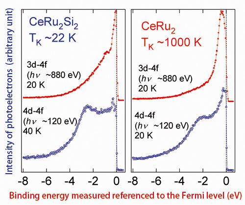
Top (red circles): Resonance spectra from the first high-resolution measurements of Ce 3d-4f. These distributions strongly reflect the bulk (inside material) electronic states. Bottom (blue circles): Ce 4d-4f resonance spectra. These distributions have been confirmed to reflect the electronic states in the surface of a material.
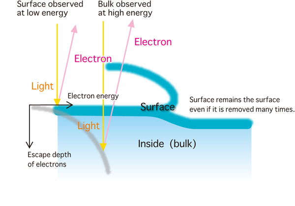
Fig. 3. Measurement principle of the bulk states within a material using soft X-ray synchrotron radiation.
Leading the World in the Development of Photoelectron Microscopes
Dr. Suga is committed to developing new measurement techniques and academic fields. In particular, a photoelectron emission microscope (PEEM), which is indispensable for research on microsized and nanosized magnetic materials, was developed through collaborative research between Dr. Suga and Dr. Jurgen Kirschner (Professor, Max-Planck-Institut für Mikrostrukturphysik, Germany). Their project, which was conducted between 1998 and 2000, demonstrated the superiority of the Soft X-ray Spectroscopy of Solid Beamline (BL25SU). BL25SU can produce perfect circular polarization of X-rays. Furthermore, Dr. Suga actively participated in the development of a spin-polarized scanning tunneling microscope (spinSTM), which should revolutionize research on nanosized magnetic materials. Moreover, he has been producing significant outcomes through collaborations with researchers at Osaka Kyoiku University, Max Planck Institute for Microstructure Physics, and Karlsruhe Institute of Technology (Germany). Through Dr. Suga’s efforts, the combined utilities of synchrotron radiation PEEM at SPring-8 and laboratory spinSTM are expected to significantly contribute to the development of new basic scientific fields.
The expectations for Dr. Suga as a leader who can extensively promote collaborative projects with Germany in research fields involving strongly-correlated electron materials and nanosized magnetic materials remain high. In addition, researchers in the field of synchrotron radiation from around the world have praised him for his dedication to encouraging young researchers to participate in these projects and mentoring world-class researchers. “I am very pleased that the cutting-edge research on photoelectron spectroscopy using synchrotron radiation at SPring-8 has been recognized. I earnestly hope that SPring-8 will continue to promote academic and cultural exchanges between young researchers around the globe and Japanese researchers as these exchanges will lead to a mutual understanding. Thereby, SPring-8 will lead the world in the advancement of science and peace,” explains Dr. Suga with gratification and hope for the future. (In 2008, Dr. Suga received the Helmholtz-Humboldt Research Award, and to this date, remains the only person in Japan to whom this honor has been bestowed upon.) The award ceremony was held in Berlin, Germany, on June 24, 2008 (see picture on the page of the list of Major Awards/Prize Winners).
Reference
1. A. Sekiyama, T. Iwasaki, K. Matsuda, Y. Saitoh, Y. Onuki and S. Suga; Nature, 403, 396 (2000)